|
Getting your Trinity Audio player ready...
|
Nonischemic cardiomyopathy (NICM) is a leading cause of heart failure and sudden cardiac death (SCD). While left ventricular ejection fraction (LVEF) has been the traditional method for risk stratification, it has limitations in identifying both high- and low-risk patients. Cardiac magnetic resonance (CMR) has emerged as a powerful tool for assessing myocardial scar, a key substrate for ventricular arrhythmias.
Late Gadolinium Enhancement (LGE) and Arrhythmic Risk Stratification
LGE is a CMR technique that detects localized areas of myocardial fibrosis, or scar tissue. Studies have consistently shown that LGE is a strong independent predictor of ventricular arrhythmias and SCD in NICM. Patients with LGE have a significantly higher risk of SCD compared to those without LGE.
Quantitative Assessment of LGE
While LGE is a qualitative technique, there are methods for quantifying the extent of LGE. Studies have shown that even small amounts of LGE can be associated with an increased risk of SCD.
LGE Location and Pattern
The location and pattern of LGE may also be important for risk stratification. For example, ring-like subepicardial LGE has been associated with a high risk of SCD in patients with desmosomal gene mutations.
T1 Mapping and Extracellular Volume Fraction (ECV)
T1 mapping is another CMR technique that can be used to assess myocardial fibrosis. ECV is a quantitative measure of interstitial fibrosis, and studies have shown that ECV is a strong predictor of ventricular arrhythmias and SCD in NICM.
CMR: A Valuable Tool for Arrhythmic Risk Stratification
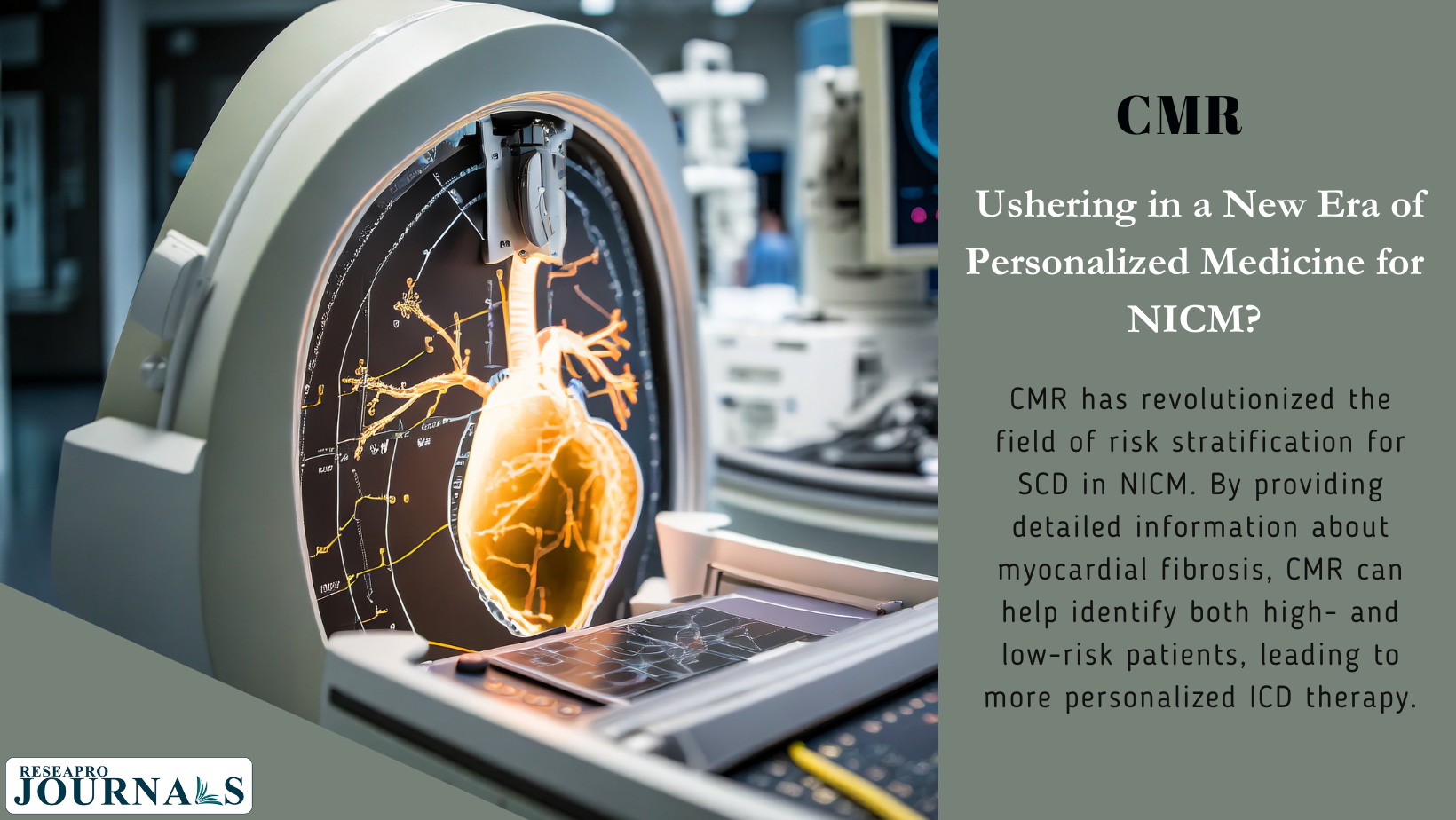
CMR is a valuable tool for arrhythmic risk stratification in NICM. LGE, T1 mapping, and ECV can provide important information about the presence and extent of myocardial fibrosis, which is a key predictor of SCD. This information can be used to guide decisions about implantable cardioverter-defibrillator (ICD) therapy. CMR has revolutionized the field of risk stratification for SCD in NICM. By providing detailed information about myocardial fibrosis, CMR can help identify both high- and low-risk patients, leading to more personalized ICD therapy.


